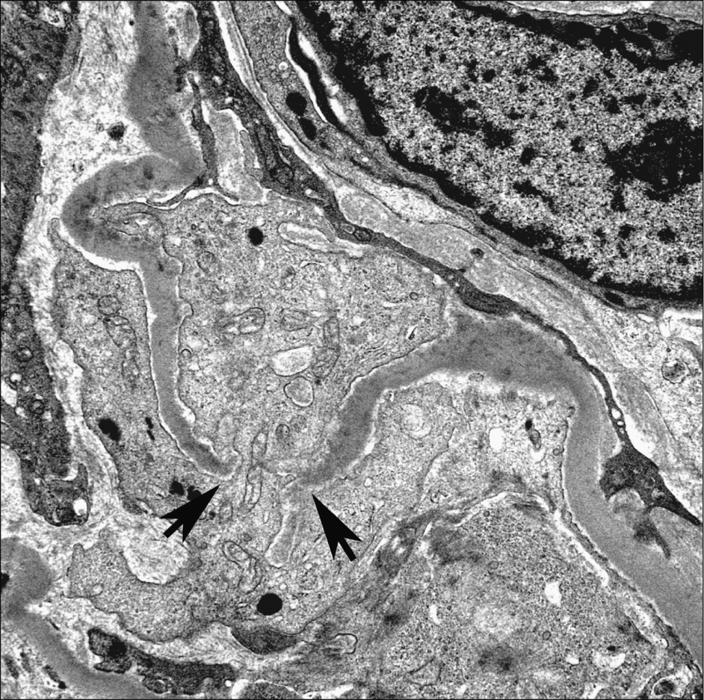
Specimens were taken from kidneys perfused with paraformaldehyde freeze fractured and then processed with conductive staining. Automatic segmentation of pathological glomerular basement membrane in transmission electron microscopy images with random forest stacks. Glomerulonephritides which develop necrotizing and crescentic lesions usually have glomerular basement membrane gbm disruptions when carefully examined by light microscopy or transmission electron microscopy.
glomerular basement membrane electron microscopy

Incidence in 3471 consecutive renal biopsies examined by electron microscopy.
Glomerular basement membrane electron microscopy. It has an electron dense lamina densa when viewed under the electron microscope and it is formed by associations among the same general classes of macromolecules found in all other types of basement membranes. This change is comparable to that seen in the heterologous phase of murine nephrotoxic nephritis. On electron microscopy such variants may produce characteristic changes within the glomerular basement membrane gbm. Archives of pathology and laboratory medicine.
2central laboratory southern medical university guangzhou 510515 china. Thin glomerular basement membrane nephropathy. Incidence in 3471 consecutive renal biopsies examined by electron microscopy. The basement membranes were shown to have a definite fine three dimentional meshwork structure composed of strands and pores resembling a childrens jungle gym the strand.
In order to clarify ultrastructural details electron microscopic observations using negative staining were performed on unfixed human and bovine glomerular basement membranes isolated by a modified method of spiro. Thin glomerular basement membrane nephropathy. Standard electron microscopy techniques have enabled the identification and classification of glomerular diseases based on two dimensional information however complex three dimensional ultrastructural relationships between. Morphological change in glomerular podocytes and the underlying basement membrane are frequently observed in disease irrespective of the underlying molecular etiology.
Despite numerous excellent and detailed ultrastructural investigations of gbm discontinuities a complete appreciation of their actual number appearance and distribution within a. Cao l1 lu y2 li c1 yang w1. These changes may be missed if glomerular lesions histologically diagnosed as fsgs on light microscopy are not. The gbm can in some ways be viewed as a typical basement membrane.
1school of biomedical engineering southern medical university guangzhou 510515 china. The fine structure of the glomerular basement membrane gbm of the rat kidney was studied by means of high resolution scanning electron microscopy. Electron microscopic studies of the kidneys of nzb nzw f 1 bw mice show the presence of electron opaque deposits on the endothelial side of the glomerular basement membrane early in the spontaneous renal disease to which these mice are subject. The fractured surface of glomerular tufts exhibited the inner and outer surface of the gbm uncovered by endothelial and.
There is an increasing appreciation that variants of the col4a genes may be associated with the development of focal segmental glomerulosclerosis fsgs.